
or red Alaskan salmon as the case may be?

moving on to the EKG…
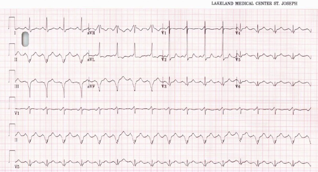
Rate sits just over 100. Rhythm appears to be sinus. Axis is left. The QRS should draw the eye somewhat. In some leads it looks narrow, in others it looks wide. In some it looks … narrow and wide. Let’s zoom in on V2.
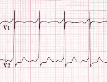
We see a short PR as well as a marked upslurring of the QRS, in other words a pretty classic delta wave. III shows the delta wave as well, though it’s inverted, consistent with the polarity of the QRS

What is the significance of finding WPW in a patient with seizures? Obviously dysrhythmia goes up as the potential cause of the seizure if the dysrhythmia made the patient hypoxic. In our patient’s case it was more of an incidental finding; it turned out the patient had a long-standing history of WPW … and then went on and had further seizure activity for us which were not associated with any dysrhythmia.
