
in honor of Halloween…

Clearly I should have run this EKG a week sooner.

Rate is borderline tachycardic. The rate is also a little irregular. Most of the RR intervals are about 3 big boxes wide so those sections are around 100, but some are wider. Counting the complexes gives an average in the low 90’s.
Rhythm was the main reason I chose to run this EKG, and it proved harder than I expected. Rhythm strips are the best places to look for P waves so let’s zoom in on the end of the V1 rhythm strip.

There’s a P before every QRS (orange dots), but did you notice the extra P wave (black dot)?
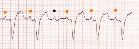
And then did you notice the extra little inflections in the ST wave? Remember the heart doesn’t depolarize during the repolarization phase, so extra inflection points there are frequently little lost P waves…

This patient is in Aflutter with mostly 2:1, and occasionally 3:1 conduction. If I’d realized how few people would spot those I’d have kept the EKG showing the flutter waves once the rate slowed down.
Moving on from the P waves, the QRS is wide with a LBBB morphology. The ST waves are appropriately discordant with the QRS. There is ST elevation especially precordially so those of you who wanted to use Sgarbossa criteria and get old EKG’s to look for changes were correct to want to assess the chronicity of the ST elevation. The lateral chest leads do have some ST depression which could be reciprocal … or could just go along with the LBBB. In this case old EKG’s did demonstrate that the ST waves and LBBB patterns were old.
Take-homes on this EKG with 2:1 and 3:1 A flutter with LBBB? First, whenever there is irregularity then one of the first questions whether Afib or flutter are present. Next, while flutter waves are frequently one big box wide, some patients (such as this one) will have slower flutter waves. Next, wide complex QRS’s can be from LBBB and don’t necessarily require a ventricular origin for the rhythm. Finally, LBBB can frequently have ST elevation, in this case an old EKG was very useful to reassure oneself that the STE was old.
One other clinical take-home, just because the patient is in A fib/flutter doesn’t mean that the tachycardia isn’t physiologic. This patient’s weakness was due to sepsis, her tachycardia was treated by managing the sepsis rather than with rate agents.
